|
||
|
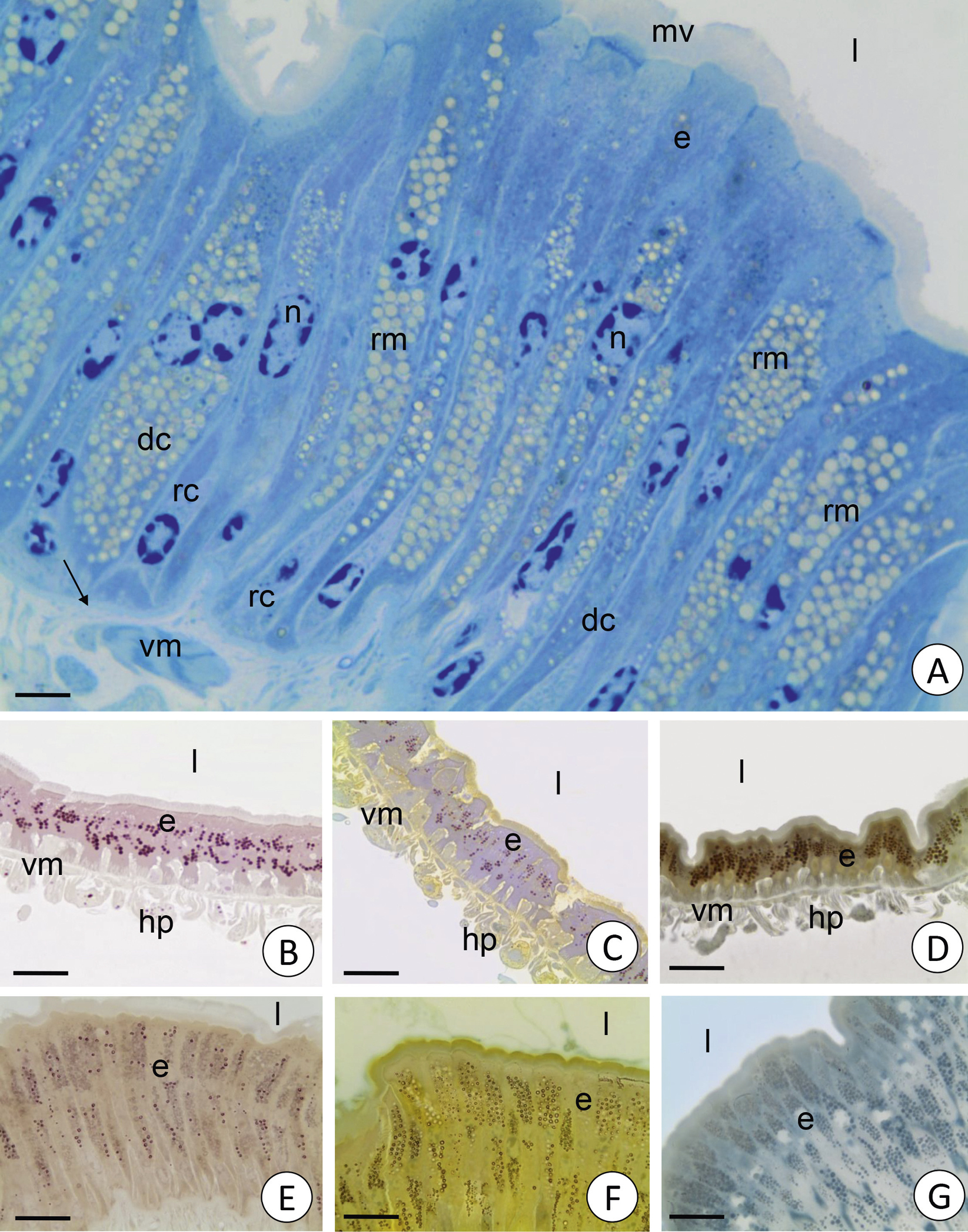
A. E. pulchripes. A pseudostratified epithelium (e) of the midgut resting on the basal lamina (arrow). Digestive cells (dc), regenerative cells (rc), midgut lumen (l), visceral muscles (vm), microvilli (mv), nuclei (n), reserve material (rm). Light microscopy. Scale bar = 5 µm. B–G. Histochemical staining of the midgut epithelium of millipedes. Midgut epithelium (e), midgut lumen (l), visceral muscles (vm), hepatic cells (hp). B. P. angustus. PAS. Light microscopy. Scale bar = 16 µm. C. P. angustus. Bonhag method. Light microscopy. Scale bar = 16 µm. D. P. angustus. Sudan Black B. Light microscopy. Scale bar = 18 µm. E. E. pulchripes. PAS. Light microscopy. Scale bar = 18 µm. F. E. pulchripes. Bonhag method. Light microscopy. Scale bar = 18 µm. G. E. pulchripes. Sudan Black B. Light microscopy. Scale bar = 12 µm. |