|
||
|
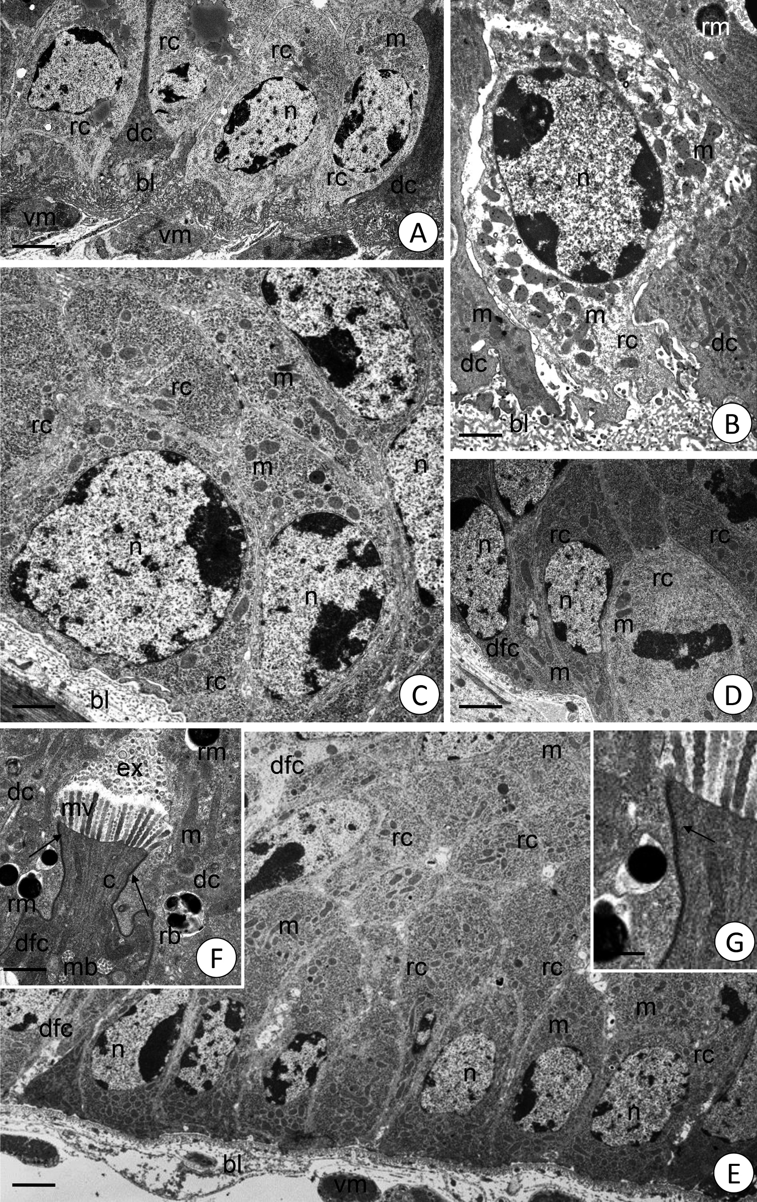
Midgut epithelia in various millipedes. A. P. angustus, transverse section. Regenerative cells (rc) among basal regions of digestive cells (dc). Nucleus (n), clusters of mitochondria (m), basal lamina (bl), visceral muscles (vm). TEM. Scale bar = 1.8 µm. B. E. pulchripes, longitudinal section. Regenerative cells (rc) among basal regions of digestive cells (dc). Nucleus (n), basal lamina (bl), clusters of mitochondria (m), reserve material (rm). TEM. Scale bar = 1 µm. C. G. tetrasticha, longitudinal section. Nest of regenerative cells (rc) in the midgut epithelium, note the cytoplasm poorly supplied with organelles. Basal lamina (bl), nucleus (n), clusters of mitochondria (m). TEM. Scale bar = 0.8 µm. D. G. tetrasticha, transverse section. Detail of a regenerative nest with dividing regenerative cells (rc) and differentiating cells (dfc), nucleus (n), mitochondria (m). TEM. Scale bar = 1.6 µm. E. G. tetrasticha. Nest of regenerative cells (rc) in the midgut epithelium displaying cytoplasm with numerous mitochondria (m). Basal lamina (bl), visceral muscles (vm), nucleus (n), differentiating cells (dfc). Transverse section. TEM. Scale bar = 1.6 µm. F. P. angustus, transverse section. A differentiating cell (dfc) with microvilli (mv) entering the extracellular space (ex). Multivesicular bodies (mb), mitochondria (m), filaments in the cortical layer (c), electron dense spheres of the reserve material (rm), residual bodies (rb), smooth septate junctions (arrows). TEM. Scale bar = 0.8 µm. G. Higher magnification of Fig. F. Smooth septate junction (arrow). TEM. Scale bar = 0.3 µm. |