|
||
|
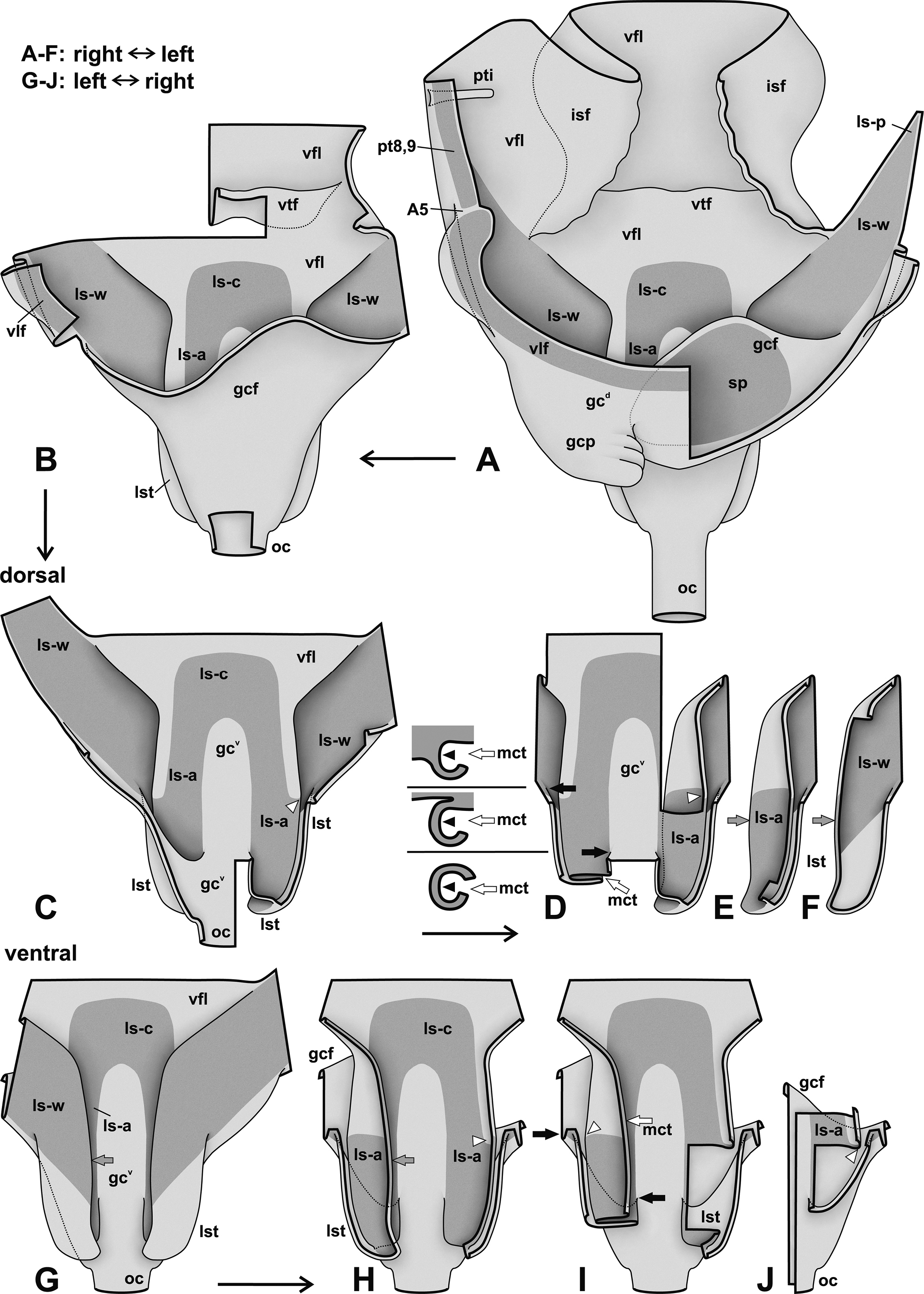
Genital chamber and laterosternal shelf area of Attaphila female, semi-schematic representation showing exoskeletal morphology, posteriorly on top. A, B: Dorsal view; selected parts removed from A to B. C–F: Dorsal view of median part of laterosternal shelf area; series of pictures with selected parts removed stepwise from C to F (only parts of one tube lst retained in E, F); lefthand of D cross sections (dorsal side up) at three anteroposterior levels shown (posterior to, in between, and anterior to the levels indicated by two black arrows in D), including mesal cleft mct of tube. G–J: Ventral view of median part of laterosternal shelf area; series of pictures with selected parts removed stepwise from G to J. — Explanations: Thick black lines are (virtual) cutting lines. Continuous thin black lines are freely visible edges (= lines along which the cuticle bends away from the observer’s view). Dashed thin black lines are edges hidden beneath other cuticle (only some shown). Membranous cuticle in very light grey, sclerotised cuticle in darker grey; cuticle shaded darker where it dives beneath other cuticle. Dashed grey lines in A show hidden part of margin of spermathecal sclerite. ― Abbreviations: A5 articulation between pt8,9 and vlf; gc genital chamber (with ventral wall gcv and dorsal wall gcd); gcf fold dividing genital chamber horizontally; gcp pouch of genital chamber; isf intersternal folds; ls laterosternal-shelf sclerite (with central part c, arm part a, wing part w, posterolateral extremity p); lst laterosternal-shelf tube (mostly sclerotised by sclerite ls: part ls-w in ventral wall; part ls-a in two further dorsal layers forming inner walls of tube); mct mesal cleft of laterosternal-shelf tube; oc common oviduct; pt8,9 extension of paratergite 9; pti paratergal invagination; sp spermathecal plate; vfl floor of vestibulum; vlf valvifer; vtf vestibular transversal fold. ― Arrows: in D and I, black arrows showing anteroposterior levels of transition between cross sections lefthand of D (corresponding to posterior end of the edge pointed to); in E, F, G, H, grey arrows indicating edge around which ls-sclerotisations ls-a and ls-w are likely continuous; in C, D, H, I, J, white arrowheads pointing to kink area of lateral border of inner tube (lst) wall where sclerotisation ls-a bends from dorsal to ventral inner wall; in sections lefthand of D, black arrowheads pointing to membranous inner lateral border of tube lst. |