|
||
|
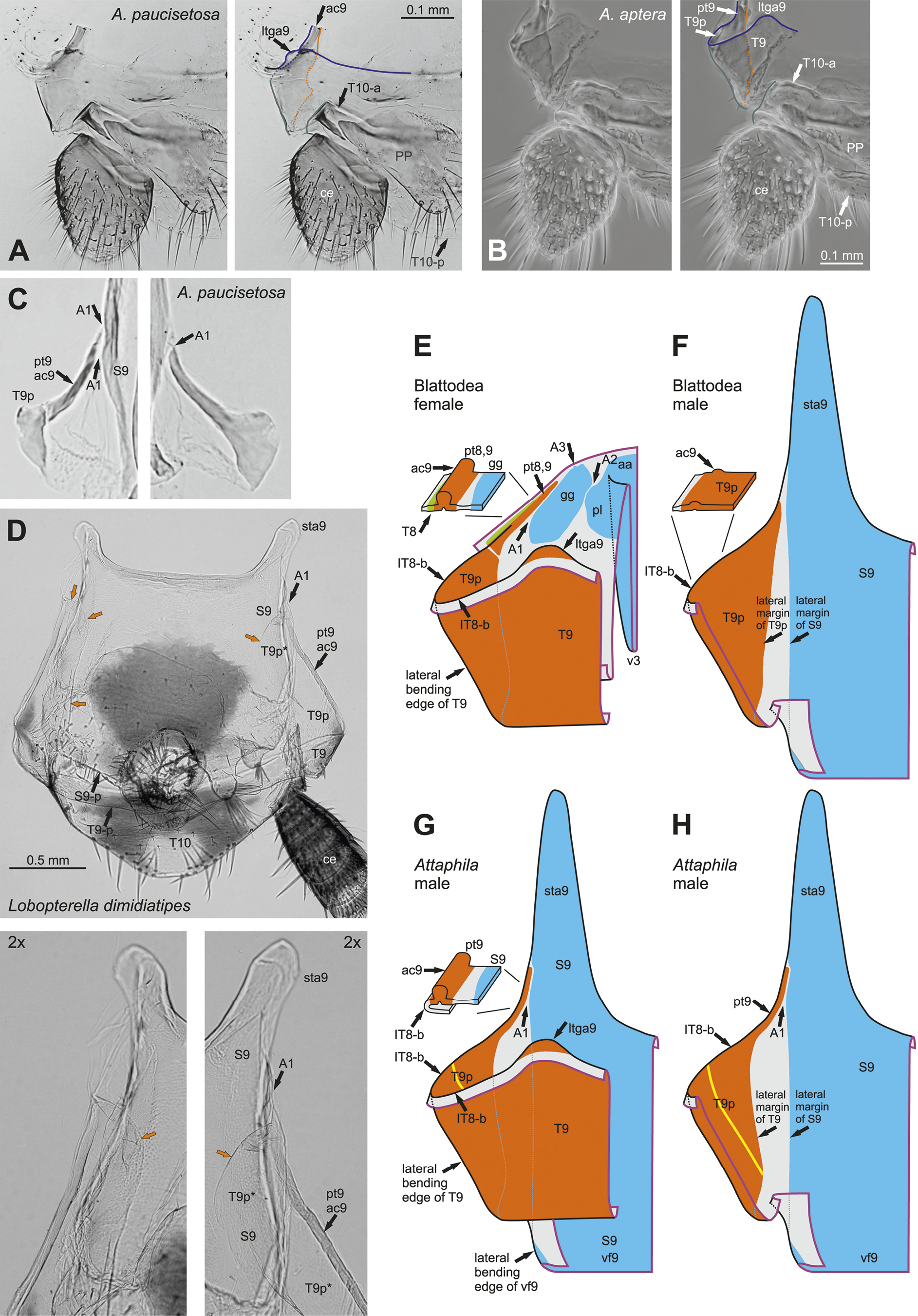
Lateral parts of tergite T9 and their relation to lateral parts of 9th-segmental (latero)coxal sclerites in males (A–D, F–H) and females (E). A: Attaphila paucisetosa (HT Bo 1258), left half of terminal abdomen, digital photograph, right picture with interpretations. B: Attaphila aptera (Bo 1256), left half of terminal abdomen, phase contrast image, right picture with interpretations. C: Attaphila paucisetosa (PT Bo 1254), parts of T9 (T9p including pt9) and S9 near their contact, for both sides of body (from Fig. 25A). D: Lobopterella dimidiatipes (ex culture), terminal abdomen (phallomeres removed), digital photograph, lower pictures showing parts of T9 (T9p including pt9; T9p* = part of T9p located underneath S9) and S9 near their contact 2× enlarged, for both sides of body; orange arrows point to ventromesal margins of T9p. E–H: Schematic drawings of left-lateral parts of segment 9 in dorsal view, with included tergal (T9, T8) and (latero)coxal (male S9; female gg, aa, pl) sclerotisations; showing generalised conditions for female (E) and male (F) blattodeans and condition in male Attaphila (G, H; dorsal parts of T9 mostly removed in H); small pictures on left top in E, F, G showing a block diagram of the lowest anterior portion of T9 (selected as shown by indicator lines). ― Abbreviations: See Supplement 1. ― Colour of lines in A, B: orange – ventrally located lateral margin of T9 (compare F, H), dashed where hidden beneath T9; blue – bending line of cuticle to the posterior (IT8-b) immediately in front of anterior margin of T9 (T9-a) (compare Fig. S1D left part); green – lateral borders of T9 and T10 (lateral bending edges where they turn to the ventral side). ― Colours and lines in E–H: Thick lines in magenta are (virtual) cutting lines through the cuticle. Continuous black lines are freely visible edges (= lines along which the cuticle bends away from the observer’s view). Dashed black lines are edges hidden beneath other cuticle (only some shown). Dashed gray lines show hidden part of lateral margins of T9p or S9. Sclerites shaded in blue (coxal and laterocoxal sclerotisations of segment 9, which together form most of “sternite” S9 in F–H), orange (tergal sclerotisations of segment 9), or green (tergal sclerotisations of segment 8); membrane shaded in light grey. Thick lines in yellow represent a potential weak zone within T9 (on part T9p). |