|
||
|
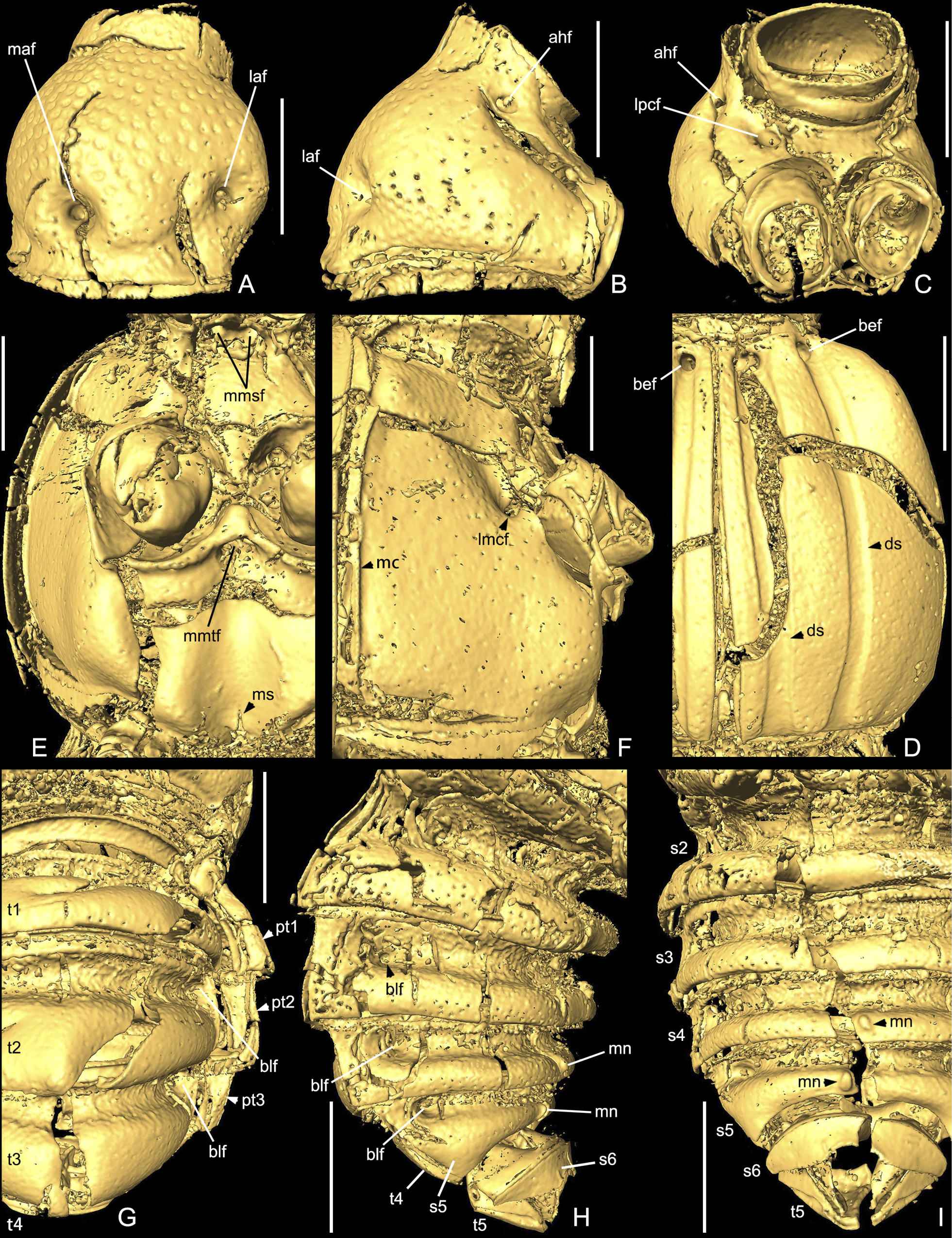
X-ray microtomographic reconstruction of Europharinodes schaufussi gen. et sp. nov., holotype, SNUC-Paleo-0102. A–C: prothorax, dorsal (A), lateral (B) and ventral (C). D Right elytron. E, F Meso- and metathorax, ventral (E) and lateral (F). G–I Abdomen, dorsal (G), lateral (H) and ventral (I). Abbreviations: ahf, antero-hypomeral fovea; bef, basal elytral fovea; blf, basal lateral fovea; ds, discal stria; laf, lateral antebasal fovea; lmcf, lateral metaventral fovea; lpcf, lateral procoxal fovea; maf, median antebasal fovea; mmsf, median mesoventral fovea; mc, marginal carina; mmtf, median metaventral fovea; mn, median nodule; ms, median split; pt1–3, paratergite 1–3; s2–6, sternite 2–6 (IV–VIII); t1–5, tergite 1–5 (IV–VIII). Scale bars: 0.2 mm. |